Following cataracts surgery, impaired vision is associated with vitreous loss. Vitreous loss is less frequent among surgeons with expertise and those who perform a considerable number of cataracts operations. Techniques for classifying patients’ risks are available to help less experienced surgeons steer clear of high-risk patients. Before having cataracts surgery, patients must be counseled of potential risks and complications as part of the therapy for vitreous loss.
The vitreous body is shielded by the posterior lens capsule, an anatomical barrier, from forces associated with intraocular lens implantation, aspiration, and fragmentation of the lens. 4.4% of patients had capsule rupture and vitreous loss in the national cataracts surgery study conducted in the UK in 1997–1998. According to other studies, patient rates might range from 8.22 to 0.45 percent. A higher risk of vision-threatening effects such endophthalmitis, retinal detachment, and cystoid macular oedema is linked to vitreous degeneration. 2, 3, 4, 5, 6, 7 An even worse prognosis for those eyes is possible if the posterior capsule is disrupted and then lens fragments are displaced into the posterior segment.
Related: How to manage the long-term complications of cataracts surgery
It is crucial for the surgeon to refrain from actions that increase the risk of an eye catastrophe when vitreous loss occurs. These include non-vitrectomy techniques to collect lens fragments from the posterior area and phacoemulsification in the presence of vitreous.
The use of sutureless 23-gauge instruments and the pars plana route, as opposed to the anterior chamber, are benefits of doing anterior vitrectomies. There is an increased risk of retinal detachment, following glaucoma, and cystoid macular oedema with lens nuclear fragment displacement into the vitreous. It is advised that a retinal surgeon treat these eyes right away.
Prevention
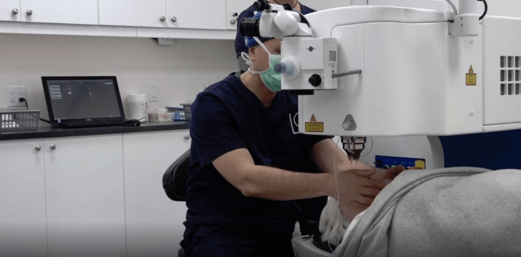
The details of safe cataracts surgery are explored in more detail in recent literature, which is beyond the purview of this article.
The amount of vitreous loss is influenced by the surgeon’s expertise, surgical volume (the number of cataracts operations each surgeon does annually),12,13, the complexity of the cases, or case mix. Prior to cataracts surgery, scoring methods allow for a measurable estimation of risk in each case. By properly preparing patients, choosing the right anesthetic, and selecting the surgeon, problems may be anticipated and prevented. In high-risk eyes, Muhtaseb demonstrated that even seasoned consultants had an 8% vitreous loss rate and a 4% incidence of falling lens nuclei. It indicates that doctors experienced in vitrectomy and the removal of misplaced lens components from the posterior area are best suited to undertake cataracts surgery in such eyes.
Although the importance of the capsulorrhexis in the success of cataracts surgery has been appropriately emphasized17, the preparation of the patient, the anesthetic used, and the design of the incision may all influence the quality of the capsulorrhexis much earlier in the procedure. In high-risk eyes, such as those that have had vitrectomy18, problems may still occur even with perfect capsulorrhexis when an unstable anterior chamber depth causes anterior chamber depth changes, pupil constriction, and patient discomfort. 19 It’s important to comprehend how to take care of these eyes.
The care of the patient
Although it is not a common treatment, individuals now anticipate having cataracts surgery. The patient will have an easier time managing a significant intraoperative problem if informed consent included topics including predicted vitreous loss, lens matter displacement, and failure to implant an intraocular lens. This will reduce patient anxiety and help the patient and physician build trust before, during, and after cataracts surgery. 93.5 percent of patients want to be told if the risk is one in 50, and 62.4 percent want to be informed if the risk is one in 1000. Patients also want to be informed about rare effects. But recall accuracy of permission information is poor, especially for serious issues, making it difficult to adequately prepare patients for potentially hazardous operations.
Management: keeping a close eye
An internal alteration in the eye that is both subtle and sudden will signal the presence of vitreous loss. The sudden appearance of a red reflex altered lens nucleus mobility, excessive sideways displacement of the nucleus, abnormal movement of structures (like the pupil margin) remote from instruments in the anterior chamber, and traction transmitted through vitreous strands are just a few symptoms that may be seen.
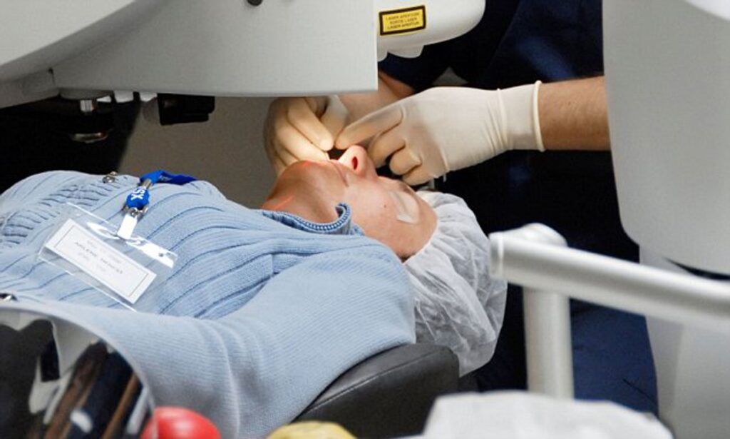
Phacoemulsification should stop if it is still going on, and the probe should be withdrawn from the eye carefully to reduce strain on the vitreous. When the phacoemulsification probe is withdrawn, a viscoelastic substance may be injected into the anterior chamber to help stabilize any remaining lens fragments and avoid vitreous prolapse.
The surgeon must then stop and meditate for a time. The vitrectomy equipment may be set up and, if required, a sub-anesthetic can be used while the situation is being evaluated. Tenon’s The lens nucleus or substantial portions of the lens material may be able to sink posteriorly if a reaction is delayed at this time. This is a problem that is controllable and often resolves well. When retrieving lens fragments using an anterior method, precipitation and force are harmful.
To finish cataracts surgery and securely implant an intraocular lens, vitreous loss management aims to remove all vitreous from the anterior chamber and operative site. Depending on the intricacy of the case and the main surgeon’s skill level, any, all, or none of these activities may be completed by the primary surgeon. A vitreous cutter and a separate infusion provided by an anterior chamber maintainer should be used to remove vitreous. Because it produces a flow conflict between infusion and aspiration and makes it difficult to maintain control of intraocular pressure, the use of a coaxial infusion sleeve around a vitreous cutter is not advised.
A drawback of so-called “dry” vitrectomy techniques is their inability to maintain a constant intraocular pressure. The cutter should be placed into the pars plana, either utilizing an anterior chamber or pars plana infusion, to collect vitreous from the anterior chamber. The vitreous may now be removed from deep beyond the posterior capsule, which is more challenging and dangerous when the cutter is put via the anterior chamber. This also decreases anterior chamber manipulation and vitreous incarceration in the corneal incision. Although triamcinolone particles may be used, their use is not necessary to view vitreous strands in the anterior chamber.
The procedure used a 25-gauge needle for sutureless vitrectomy. The bigger 23-gauge sutureless devices provide more control and could be more successful in removing vitreous as well as retained lens debris. Soft lens debris may easily be removed using the vitrector.
The nuclear material may either be emulsified and aspirated by reintroducing the phacoemulsification probe or intact nuclear fragments can be removed through an enlarged incision once the anterior chamber has been completely cleared of vitreous. Nuclear fragments may be stabilized with viscoelastics or a lens glide29 before to removal from the anterior chamber.
Forceful efforts to remove nuclear fragments without a pars plana vitrectomy may cause significant retinal tears and detachment if they are moved posteriorly past the plane of the posterior capsule.